VAGINITIS CASE 1
ANSWER 4
Wet preps are
obtained using a Q-tip applied to the vaginal side-wall within the largest pool
of discharge. Drop the Q-tip with the sample of discharge into a test tube
with approximately 2 ml of normal saline (not water) in it. A drop of the
suspension is then placed on a slide, covered with a cover-slip, and carefully
examined.
Under low power, observe for:
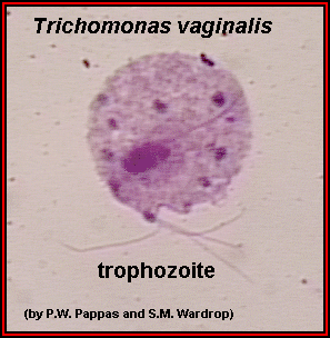
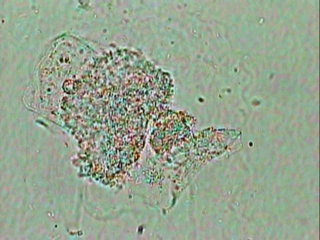
- Trichomonads –

are motile pear-shaped organisms with active flagella,
larger than a WBC but smaller than epithelial cells, that usually are seen
swimming or thrashing around in the wet prep. - WBC – presence and number of white blood cells. Lots
of PMNs indicates inflammation which is seen more with trichomoniasis,
cervicitis, and to some degree with atrophic vaginitis. - Round or oval parabasal cells – normal vaginal squames are
rectangular or sharp-edged, but an atrophic vagina sheds round or oval
cells.
Under high power, observe for:
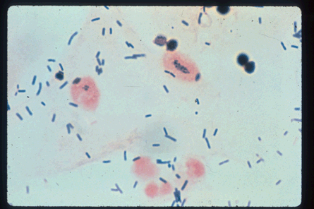
- Clue cells

these are epithelial cells that have tiny pleomorphic bacteria adherent to
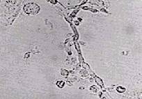
their surfaces, obscuring their borders and causing a stippled appearance. - Yeast or hyphae

- Lactobacilli – normal
flora.
KOH prep is made
by adding a drop of 10% KOH solution to a drop of saline suspension of the
discharge. The KOH lyses epithelial cells in 5 minutes (faster if the slide is
warmed briefly over a flame) and allows easier microscopic visualization of
Candidal hyphae. Other fungal stains may also be used. Swartz-Lamkin
stain contains a base for lysis of epithelial cells and india ink, which is
taken up by fungal cells.
Whiff test may be
done either by adding a few drops of KOH to the vaginal discharge remaining on
the lip of the speculum or by sniffing the KOH slide just after the KOH is
added. A foul, fishy odor is indicative of anaerobic overgrowth and, thus,
bacterial vaginosis (though trichomonas can cause false positivies).
pH test is
performed by dipping pH paper into the discharge remaining on the vaginal
speculum (before doing the whiff test)
